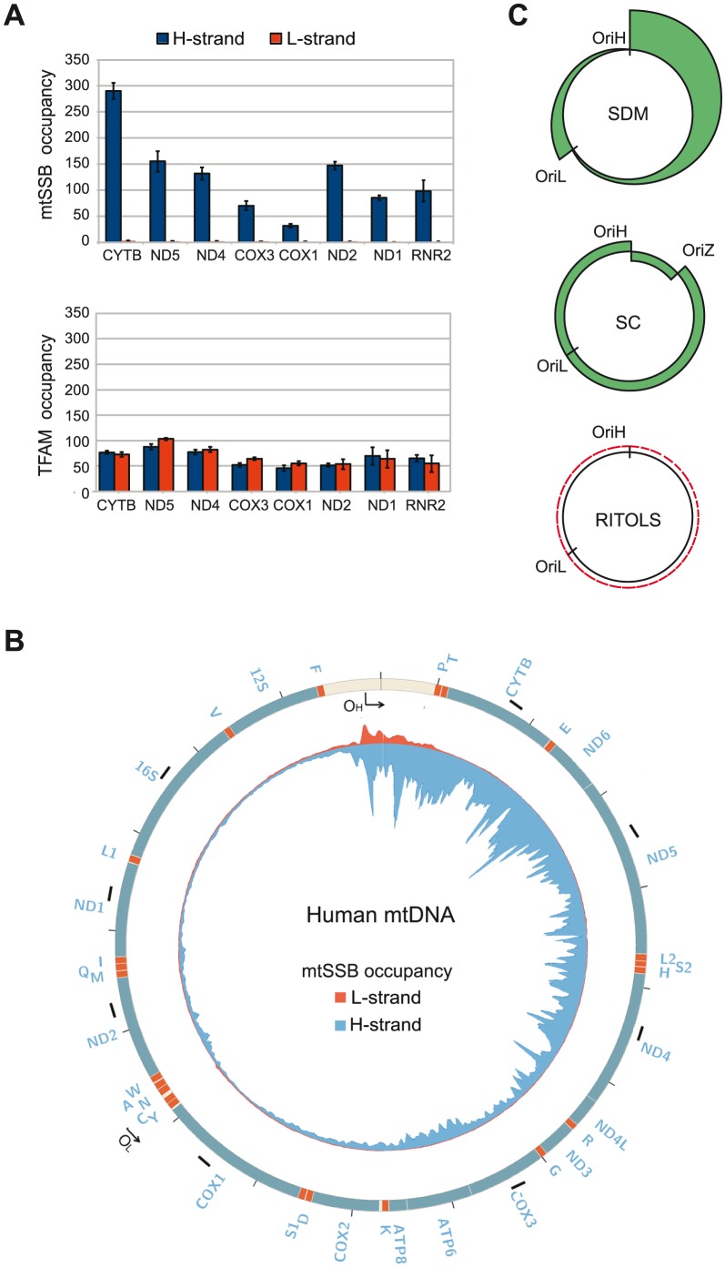
Figure 4. mtSSB in vivo occupancy reflects strand-displacement mtDNA replication.
(A) Occupancy of mtSSB and TFAM analyzed by strand-specific qPCR amplification of ChIP samples. (B) Strand-specific ChIP-seq profile of mtSSB binding to mtDNA. The origins of replication are indicated. The short black bars indicate the location of the primers used for strand-specific qPCR. (C) Schematic illustration of expected occupancy of mtSSB accordingly to the different mtDNA replication models. SDM (Strand displacement mode), SC (Strand coupled mode), and RITOLS.

