
Figure 6. aldh1a3 is required to pattern the zebrafish otic vesicle.
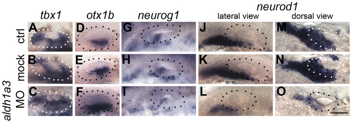
(A–C) Control uninjected (A, n = 36) and mock-injected (B, n = 14) embryos show normal expression of tbx1. Embryos injected with the aldh1a3 morpholino (C) show an expansion of expression of tbx1 in the anteroventral part of the OV (n = 38/46). (D–F) Control uninjected (D, n = 98) and mock-injected (E, n = 25) embryos show normal expression of otx1b. Embryos injected with the aldh1a3 morpholino (F) show a marked expansion of otx1b expression in the anteroventral part of the OV (n = 95/122). (G–I) Control uninjected (G, n = 11) and mock-injected (H, n = 9) embryos show normal expression of neurog1. Embryos injected with the aldh1a3 morpholino (I) show a marked reduction of neurog1 expression in the OV (n = 33). (J–O) Control uninjected (J,M, n = 42) and mock-injected (K,N, n = 18) embryos show normal expression of neurod1. Embryos injected with the aldh1a3 morpholino (L,O) show a marked reduction of neurod1 expression (n = 70/76). (A–L) Lateral views; (M–O) dorsal views; anterior to the left. Scale bar: 50 µm.