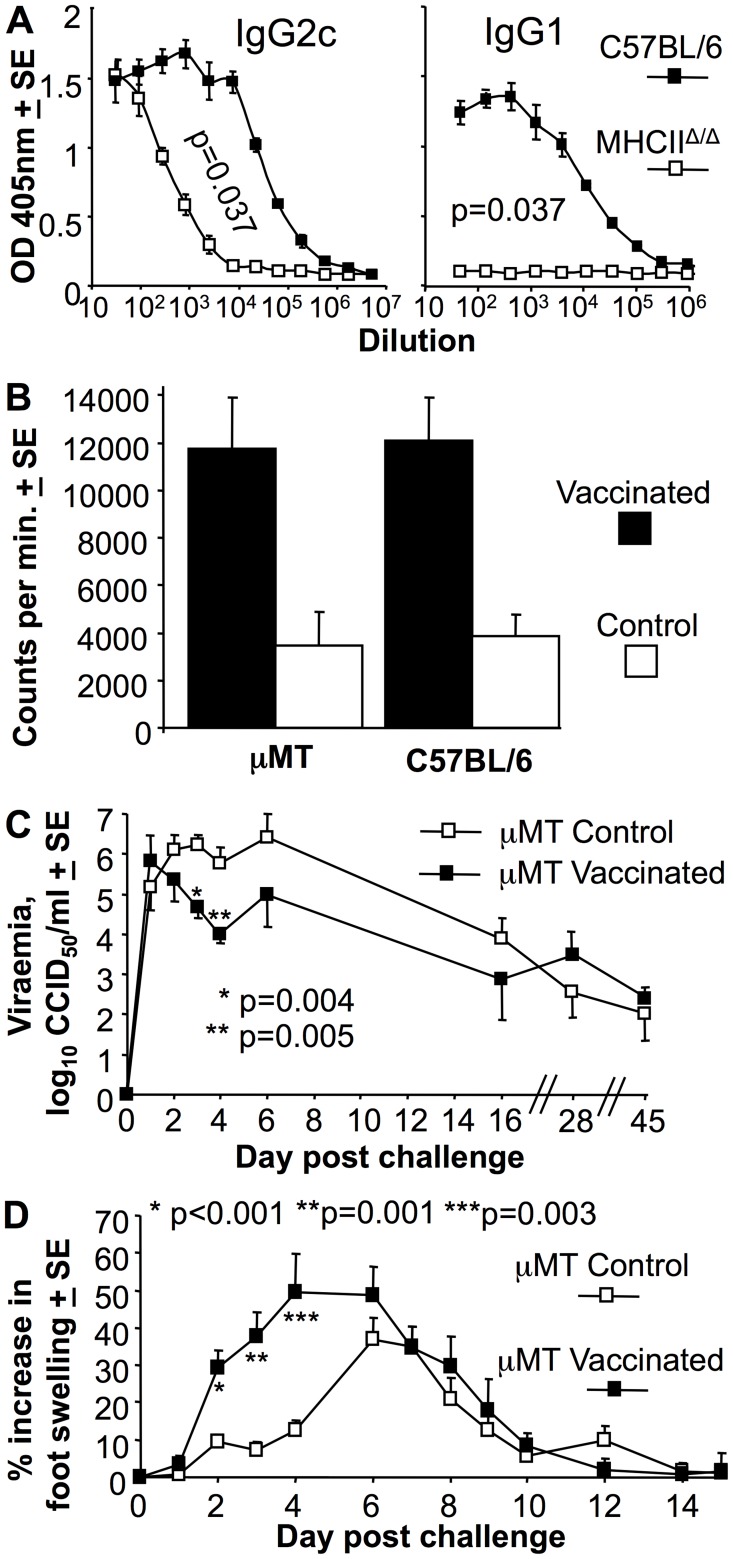
Figure 2. MHCIIΔ/Δ mice generate IgG2c responses and vaccinated µMT mice show lower early viraemia, but exacerbated arthritic disease.
(A) MHCIIΔ/Δ and C57BL/6 mice were analyzed for CHIKV-specific IgG2c and IgG1 levels by ELISA at 21 days post-infection (n = 4 per group). Statistics by Kolmogorov-Smirnov test comparing 50% end point titers. (B) µMT and C57BL/6 mice were inoculated with 10 µg of inactivated CHIKV vaccine (Vaccinated) or PBS s.c. (Control). Splenocytes were harvested day 21 post infection and used in a standard proliferation (tritiated thymidine incorporation) assay using inactivated CHIKV as antigen. Shown is the mean of 2 independent experiments both of which showed significant differences between vaccinated and Control for each mouse strain (µMT p = 0.013 and 0.043; C57BL/6 p = 0.037 and 0.027, statistics by Kolmogorov-Smirnov and t test (n = 4–5 per group). (C) Viraemia following standard CHIKV challenge of µMT mice that had received PBS (µMT Control) or 10 µg of inactivated CHIKV vaccine s.c. (µMT vaccinated) 3 weeks previously. Statistics by Mann Whitney U or Kolmogorov-Smirnov tests, (n = 6–10 mice per group; data from 2 independent experiments). (D) Foot swelling following standard CHIKV challenge of µMT mice that had received PBS or CHIKV vaccine as for C. Statistics by Kolmogorov-Smirnov tests (n = 8–20 feet per group; data from 2 independent experiments).

