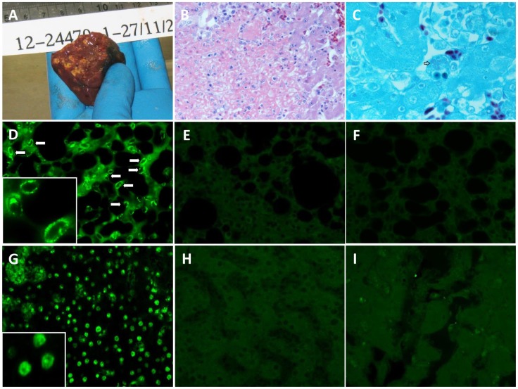
Figure 1. Illustrative histopathological changes of infected birds.
Panels A–C: Macroscopic and microscopic changes in the liver of bird 1. Panel A: cut surface of liver showing numerous 1–2 mm coalescing necrosis. Panel B: areas of acute coagulative necrosis, which are randomly distributed throughout the liver (hematoxylin and eosin staining; original magnification, ×400). Panel C: Intracytoplasmic accumulations of Chlamydial organisms (arrow) are commonly found throughout the liver parenchyma (gimenez staining; original magnification, ×1000). Panels D–I: immunochemical staining for Psittacine adenovirus HKU1 fiber protein (Original magnification, ×400). The tissue sections were deparaffinized and rehydrated, followed by blocking with 1% bovine serum albumin in PBS to minimize non-specific binding. The sections were incubated with immune serum (panels D, F, G, I) or with non-immune serum (panels E and H) of guinea pigs. After washing three times with PBS, the sections were incubated with FITC-conjugated rabbit anti-guinea pig IgG. For bird 2, apple green fluorescent foci were seen in the cytoplasm of pneumocytes (D) and in the nucleus of hepatocytes (G) with immune serum. As controls, fluorescent foci were not detected in the lung (E) and liver (H) tissue of bird 2 with non-immune serum. For bird 3, fiber protein-positive cells were not detected in the lung (F) and liver (I) tissue.

