FIGURE 3.

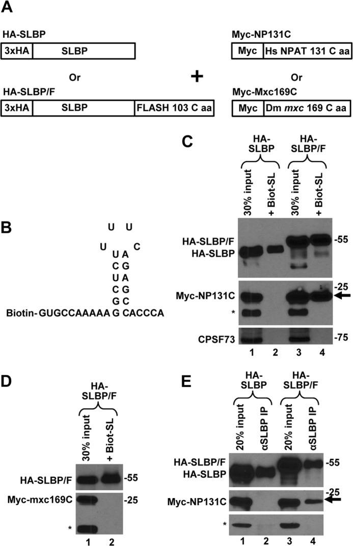
C-terminal regions of FLASH and NPAT interact in HeLa cells. A, schematic representation of clones transiently expressed in HeLa cells. B, nucleotide sequence of the Biot-SL RNA used to purify SLBP. C, Myc-NP131C was co-expressed in HeLa cells with either HA-SLBP (lanes 1 and 2) or HA-SLBP/F (lanes 3 and 4). SLBP was affinity-purified by the Biot-SL RNA from whole cell lysates prepared from transfected HeLa cells and the precipitated material tested for the presence of the transiently expressed HA- and Myc-tagged proteins (lanes 2 and 4). Lanes 1 and 3 contain 30% of the material used for purification. Myc-NP131C co-purified by virtue of interacting with HA-SLBP/F is indicated with an arrow. A protein cross-reacting with the anti-Myc antibody (indicated with an asterisk) served together with CPSF73 as a control to measure the extent of nonspecific background present in the purified material. D, Myc-Mxc169C and HA-SLBP/F were co-expressed in HeLa cells and the interaction between these two proteins tested as described in C. A protein cross-reacting with the anti-Myc antibody is indicated with an asterisk. E, Myc-NP131C was co-expressed in HeLa cells with either HA-SLBP (lanes 1 and 2) or HA-SLBP/F (lanes 3 and 4). SLBP was precipitated by an anti-SLBP antibody (αSLBP) and the precipitated material tested for the presence of the transiently expressed HA- and Myc-tagged proteins (lanes 2 and 4). A protein cross-reacting with the anti-Myc antibody is indicated with an asterisk, and the co-precipitated Myc-NP131C is indicated with an arrow.
