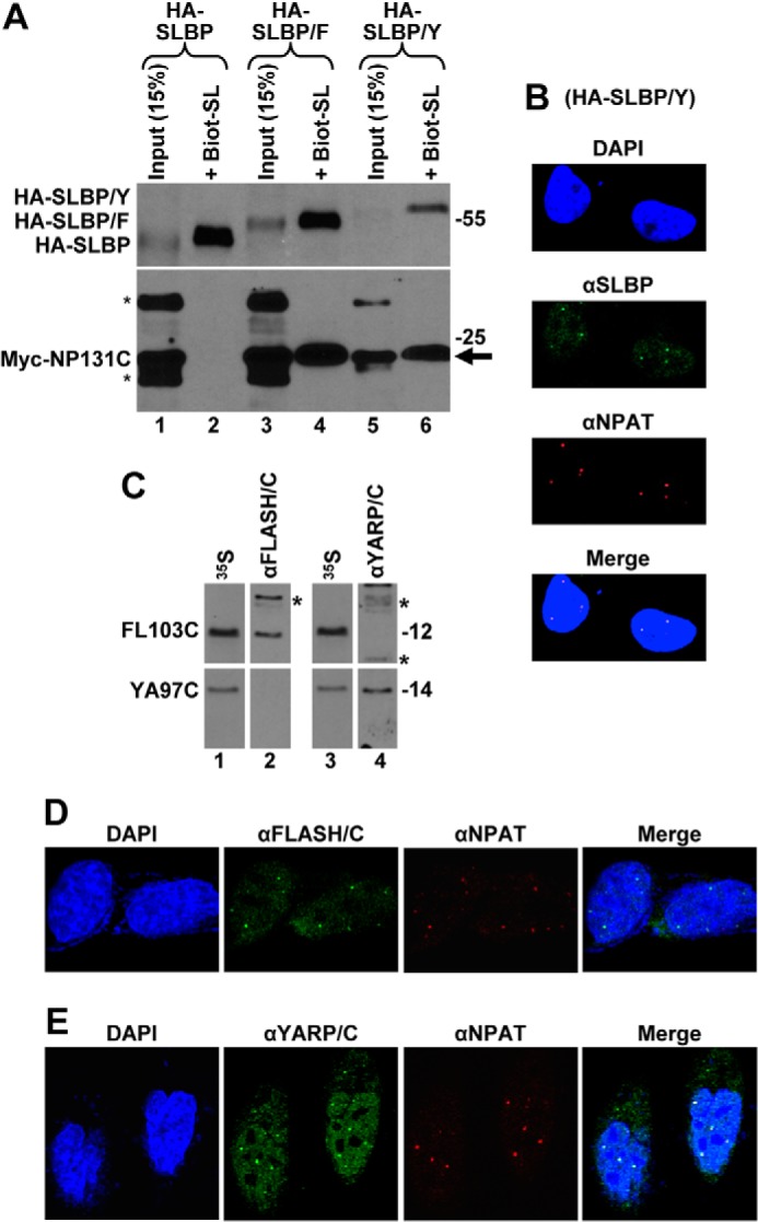
FIGURE 6.

YARP is a component of HLBs in HeLa cells. A, Myc-NP131C was co-expressed in HeLa cells with either HA-SLBP (lanes 1 and 2), HA-SLBP/F (lanes 3 and 4), or HA-SLBP/Y (lanes 5 and 6). SLBP was affinity-purified by the Biot-SL RNA from whole cell lysates prepared from transfected HeLa cells, and the precipitated material was tested for the presence of the transiently expressed HA- and Myc-tagged proteins (lanes 2, 4, and 6). Lanes 1, 3, and 5 contain 15% of the material used for purification. Co-purified Myc-NP131C is indicated with an arrow, and proteins cross-reacting with the anti-Myc antibody are indicated with asterisks. B, fraction of SLBP in HeLa cells transiently expressing HA-SLBP/Y localizes to HLBs. SLBP was detected by an anti-SLBP antibody and stained green. HLBs were detected using the DH3 mouse monoclonal antibody (αNPAT) and stained red. C, rabbit antibodies targeted to the C-terminal regions of FLASH or YARP are free of cross-reacting activities. FL103C and YA97C were expressed in rabbit reticulocyte lysate and labeled by incorporating [35S]methionine. Duplicate samples of each labeled protein were separated in an SDS-polyacrylamide gel, transferred to a nitrocellulose membrane, detected by autoradiography (lanes 1 and 3), and subsequently immunoblotted with the cross-adsorbed αFLASH/C (lane 2) or αYARP/C (lane 4) antibodies. Proteins of the rabbit reticulocyte lysate that cross-react with each antibody are indicated with asterisks. D and E, endogenous FLASH (D) and YARP (E) were detected in HeLa cells by αFLASH/C and αYARP/C antibodies, respectively, and stained green. HLBs were detected by an antibody against NPAT and stained red. Nuclei were visualized by staining with DAPI.
