FIGURE 2.
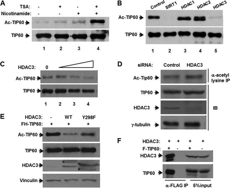
TIP60 interacts with and is deacetylated by HDAC3. A, HEK293 cells were transfected with pTOPO-FLAG-TIP60 for 24 h and then incubated with or without 1 μm TSA and/or 5 mm nicotinamide, as indicated, for an additional 6 h. An in vivo acetylation assay (Ac-TIP60) and Western blot analysis were then performed. B, HEK293 cells were transfected with FLAG-TIP60 alone or with deacetylases as indicated. Deacetylation assays were then performed. C, HEK293 cells were transfected with FLAG-TIP60 alone or with increasing amounts of HDAC3 constructs, followed by a deacetylation assay. D, H1299 cells were transfected with luciferase siRNA or HDAC3 siRNA using Lipofectamine 2000 reagent. Whole cell lysates were subjected to anti-acetyl lysine IP. The immunoprecipitates and whole cell lysates were then analyzed by Western blotting with the indicated antibodies. IB, immunoblot. E, HEK293 cells were transfected with FH-TIP60 alone or with HDAC3 or F-HDAC3-Y298F, followed by a deacetylation assay. The asterisks indicate nonspecific bands. F, HEK293 cells were transfected with the indicated plasmids. Whole cell lysates were subjected to M2 bead IP, and the resulting immunoprecipitates and 5% input were analyzed by antibodies against HDAC3 and TIP60.

