FIGURE 1.
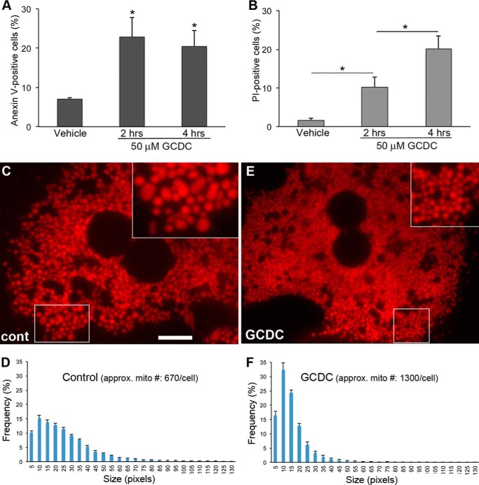
Toxic bile salt GCDC induces cell death and mitochondrial fragmentation in primary mouse hepatocytes. A and B, primary mouse hepatocytes were incubated with 50 μm GCDC for the indicated time, and cell death was evaluated using annexin V (A) and PI (B) staining. 100–200 cells were counted in each treatment; the experiment was repeated three times. Error bars represent S.E. *, p < 0.05. C—F, GCDC-induced mitochondrial fragmentation. Spherical/ovoid mitochondria in primary hepatocytes under control conditions (C) became smaller after GCDC treatment (E). Scale bar: 10 μm. Insets: 2× magnified. Images were acquired after 2 h of GCDC incubation. Frequency plots for mitochondrial size show a broader distribution of different sizes of mitochondria under control conditions (D) versus smaller and more uniform mitochondria with an ∼2-fold increase in number after GCDC treatment (F).

