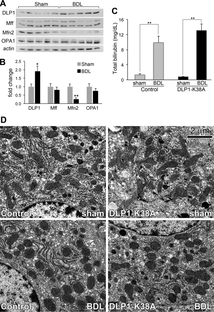
FIGURE 7.
Mitochondrial morphology in the livers after bile duct ligation. A and B, fission and fusion proteins in livers from sham- and BDL-operated mice (5 mice each) were analyzed by immunoblotting (A). Quantification indicates a significant increase of DLP1 and a marked decrease of Mfn2 in BDL mice (B). C, increased serum total bilirubin in both control and DLP1-K38A livers after BDL. Error bars represent S.E. *, p < 0.05; **, p < 0.01. D, electron micrographs of livers from control and tTg (DLP1-K38A) at 3 days post BDL. Mitochondria are elongated in sham-operated livers (Control sham, DLP1-K38A sham), whereas they appear fragmented in BDL livers (Control BDL, DLP1-K38A BDL).

