FIGURE 9.
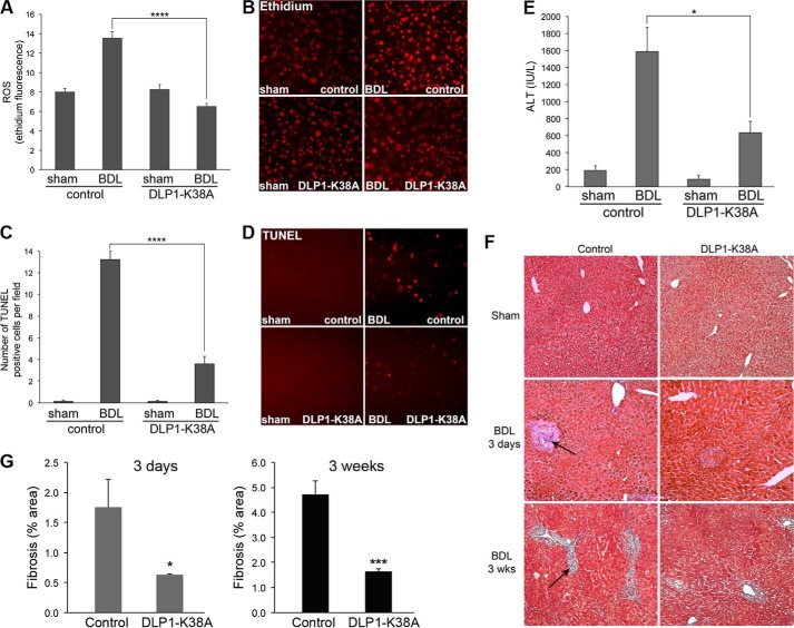
DLP1-K38A expression decreases ROS levels, apoptosis, and fibrosis in cholestatic livers. A–E, BDL for 3 days increases ROS levels (A, B), apoptosis (C, D, TUNEL), and liver injury (E, ALT) in control mice (n = 5). Livers from DLP1-K38A mice 3 days after BDL show significant decreases in these parameters (n = 5). *, p < 0.05; ****, p < 0.0001. F, trichrome-stained sections from 3 days and 3 weeks of BDL liver. Control mice show a significant increase in fibrosis (bluish fibrous staining, arrows) after 3 days, which is more pronounced after 3 weeks. DLP1-K38A expression significantly reduces fibrosis at both 3 days and 3 weeks post-BDL. G, quantification of liver fibrosis. At 3 days post BDL, n = 5 for both control and DLP1-K38A mice; at 3 weeks post-BDL, n = 3 for control mice and n = 5 for DLP1-K38A mice. *, p < 0.05; ***, p < 0.0001.

