FIGURE 11.

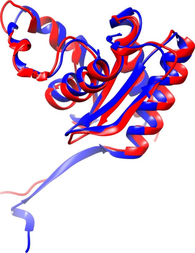
Structural alignment of one chain of HbpS from S. reticuli (blue; PDB code 3FPV) superimposed to 2.5 Å over 125 residues to OrfY from Klebsiella pneumoniae (red; PDB code 2A2L) calculated with SALAMI (69).

Structural alignment of one chain of HbpS from S. reticuli (blue; PDB code 3FPV) superimposed to 2.5 Å over 125 residues to OrfY from Klebsiella pneumoniae (red; PDB code 2A2L) calculated with SALAMI (69).