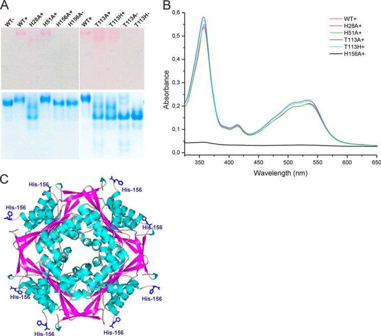
FIGURE 7.
HbpS uses His-156 to bind H2OCbl+. 20 μm concentrations of either HbpS wild type apoprotein or its mutant versions were incubated with 80 μm H2OCbl+ (WT+, H28A+, H51A+, H156A+, T113A+, and T113H+). As a control, four samples were incubated in buffer lacking H2OCbl+ (WT−, H156−, T113A−, and T113H−). The mixtures (containing 10 μg of each protein) were either loaded onto a native PAA gel (A) or analyzed by UV-visible spectroscopy after gel filtration (B). The native gel was scanned after electrophoresis (A, top) and subsequently stained with PageBlue (A, bottom). C, the exposed His-156 (in blue) on the surface of the HbpS octamer (PDB code 3FPV) is shown.

