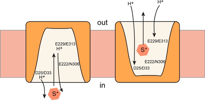
FIGURE 7.

A simplified view of the catalytic cycle of BbMAT and rVMAT2. For the sake of simplicity, the schematic includes only the conformations where the protein faces the inside (in) or the outside (out), with two protons moving to the inside of the cell in the case of BbMAT or to the vesicular lumen in the case of rVMAT2. In the schematic, the conserved amino acids in BbMAT and the corresponding ones in rVM AT2 are shown, and their hypothetical role in substrate and proton binding is presented.
