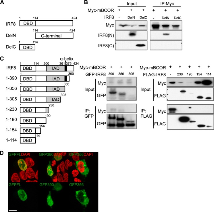
FIGURE 3.
BCOR interaction with the C terminus of IRF8 and nuclear localization. A, diagram of N and C termini-deleted IRF8 constructs. DBD, DNA binding domain. B, HEK293 cells expressing N (DelN) and C termini deleted (DelC) IRF8s and Myc-tagged full-length BCOR were lysed, pulled down with anti-Myc antibody, and subsequently blotted for BCOR and both the N-terminal and C-terminal IRF8s. The C terminus-deleted IRF8 failed to co-immunoprecipitate with Myc-mBCOR. IRF8(N), antibody raised against the N-terminal region of IRF8; IRF8(C), antibody raised against the C-terminal region of IRF8. C, diagram of a series of C termini-truncated GFP/FLAG-IRF8 constructs. DBD, DNA binding domain (white box); IAD, IRF association domain (gray box); α-helix, α-helix conserved region (black box). HEK293 cells expressing a series of C termini-truncated GFP/FLAG-IRF8 and Myc-tagged full-length BCOR were lysed, pulled down with anti-GFP or anti-FLAG antibody, and subsequently blotted for GFP, FLAG, and Myc. BCOR was co-precipitated with 390 IRF8 that only contains the α-helix region. D, images of C termini-truncated GFP-IRF8 (green)-transfected HEK293 cells. Nuclei were counterstained with DAPI (red). GFPFL, GFP-full-length IRF8 (amino acids 1–424); GFP390, GFP-IRF8 (amino acids 1–390); GFP356, GFP-IRF8 (amino acids 1–356). Scale bar, 100 μm. IP, immunoprecipitation.

