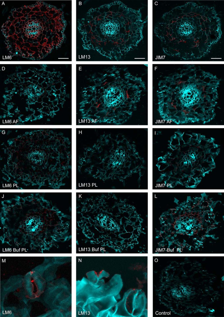
FIGURE 8.
Epitope detection of spinach root and leaf sections. Shown are confocal microscopy images of fixed spinach root sections either untreated (A–C) or treated with AF (D–F), PL (G–I), or PL buffer (Buf) (J–L) and then probed with antibody LM6 (all arabinose; A, D, G, and J), LM13 (arabinan; B, E, H, and K), or JIM7 (homogalacturonan; C, F, I, and L). Spinach leaves were probed with LM6 (M) or LM13 (N). Root and leaf tissues were stained with Calcofluor white (light blue), and antibodies were detected with the secondary Alexa Fluor 568-conjugated antibody (red). Scale bars, 8 μm.

