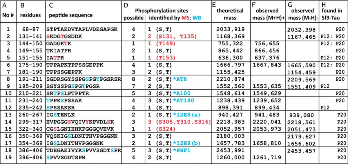
TABLE 1.
Phosphorylated peptides of Sf9-Tau (P20 and P12) observed by mass spectrometry
Column A, tryptic peptides were obtained from P12-Tau and P20-Tau expressed in Sf9 cells and analyzed by MALDI-TOF-MS; the table lists 19 phosphorylated peptides. Column B, residue numbers of each peptide (start/end) are according to the hTau40 sequence. Column C, amino acid sequences of the identified peptides with highlighted residues are shown. Red letters are for MS-identified phosphorylated residues, and blue letters are for phosphorylation sites identified by phosphorylation-specific antibodies. Column D, possible number (left column) and number of identified (right column) phosphorylation sites in the peptides are shown. If the exact site could not be identified by MS (red) and/or Western blot (WB, blue), only the number of phosphates is given with the possible residues (S, serine; T, threonine). Column E, theoretical mass was calculated for the peptide. Column F, observed mass of peptide at the positive reflector ([M + H]+) is shown. Column G, observed mass of peptide at the negative reflector ([M − H]−) is shown. Column H lists the phosphorylated peptides found in samples of P12-Tau and/or P20-Tau.

