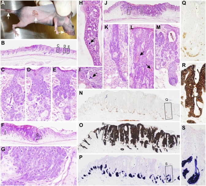
Figure 1. Cutaneous papillomas and trichoblastomas in mice.
A representative B6.Cg-Foxn1nu / Foxn1nu (nude) mouse was scarified and inoculated with MmuPV1 on the muzzle, dorsal, and tail skin. Verrucous proliferations arose at each of the injection sites (A, arrows). The earliest lesions consisted of acanthosis, small focal ulcers, and medusiform proliferations of hair follicle root sheaths around and above the sebaceous glands (B–E). Slightly more advanced lesions had larger sheets of basaloid cells with hyperchormatic nuclei and a high mitotic index (F, G) occasionally with tripolar mitotic figures (arrows, H, I). Other invasive lesions (J) had long extensions from the base of hair follicles (K, enlargement of boxed area in J), proliferative areas with “keratin” pearl formations (arrows, L) or normal early-stage anagen follicles with medusiform proliferating root sheath cells in the bulge area resulting in concurrent follicular dystrophy (M). Sebaceous glands were unaffected as reserve cells were not expanded nor in abnormal locations when labeled for SOAT1 expression (N, Q, enlargement of boxed area in N), were completely outlined by the proliferating root sheath cells based on KRT14 expression (O, R, enlargement of boxed area in O), and mature sebocytes were organized in a normal proliferation pattern when labeled with adipophilin (P, S, enlargement of boxed area in P).

