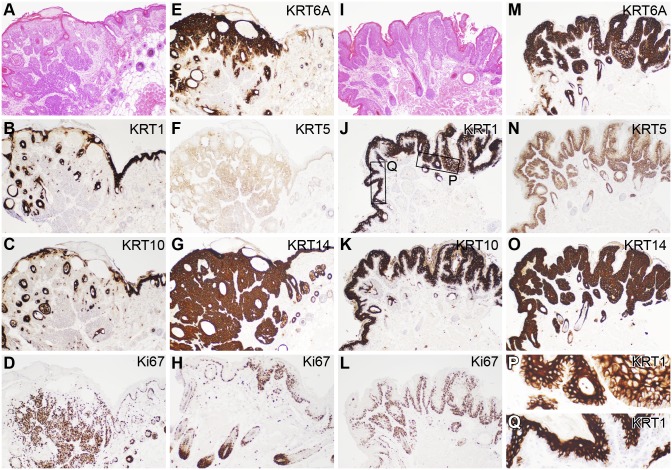
Figure 5. Immunohistochemical comparison of mouse trichoblastoma and papilloma.
Trichoblastomas exhibited both epidermal proliferation and irregular invasion into the dermis and hypodermal fat layers (A, H&E stain) in comparison to papillomas that were exophytic (I, H&E stain). KRT1 and 10, normally expressed in the suprabasal epidermis, were downregulated in the trichoblastomas (B, C) but upregulated in the papillomas (J, K). However, in papillomas, while these keratins were suprabasalar in expression in normal and hyperplastic epidermis, their expression was throughout the epidermis in the papillomas (P, Q). KRT5 and 14, normally found primarily in basal cells, were diffusely upregulated in the trichoblastomas (F, G) as well as the papillomas (N, O), although KRT5 was more localized to expressed in basal cells of papillomas. Ki67, a marker of cell proliferation, was diffuse in the invading trichoblastoma (D) in sharp contrast to the junction of the trichoblastoma and hyperplastic epidermis (H) or papillomas (L) where expression was primarily restricted to basal cells and anagen bulbs.

