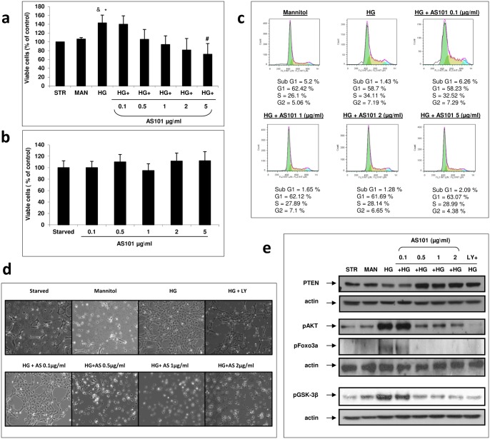
Figure 4. AS101 attenuates high glucose induced mesangial cell proliferation.
Mesangial cells were cultured in DMEM without serum for 24 h. Cells were then transferred to fresh DMEM without serum in the presence or absence of HG treatment (30 mM). For osmotic control, cells were treated with Mannitol (24.5 mM). Medium for the serum deprived control (Starved) or the osmotic control (Mannitol) contained normal levels of glucose (5.5 mM). (a) Cells treated with HG were supplemented with different concentrations of AS101 (0.1, 0.5, 1, 2, 5 µg/ml). After 24 h, XTT assay was performed. OD levels were compared to control which was normalized to 100%. Results represent mean±SEM from three experiments. #p<0.05 decrease vs. HG. *p<0.05 increase vs. starved. &p = 0.057 increase vs. mannitol. (b) Cells treated with Normal glucose levels were supplemented with AS101 at the concentrations indicated in panel a. After 24 h, XTT assay was performed. OD levels were compared to control which was normalized to 100%. Results represent three experiments. (c) Cells treated with HG were treated with different concentrations of AS101 (0.1, 1, 2, 5 µg/ml). After 48 h of treatment, cells were stained with PI buffer; cell cycle analysis was performed by FACS to determine the level of cell accumulation in each cell cycle phase: sub-G1, G1, S, G2. Results represent two experiments. (d) Cells treated with HG were treated with different concentrations of AS101 (0.1, 0.5, 1, 2 µg/ml) or LY294002 (50 µM). After 24 h, mesangial cell expansion was examined by light microscopy (X40). Results shown are from a single experiment representative of seven. (e) Cells treated with HG were treated with different concentrations of AS101 (0.1, 0.5, 1, 2 µg/ml) or LY294002 (50 µM). After 24 h, cell lysates were extracted for detection of PTEN, p-AKT, p-GSK3β, p-FoxO3a by western blot analysis, with actin as loading control. Results represent three experiments.

