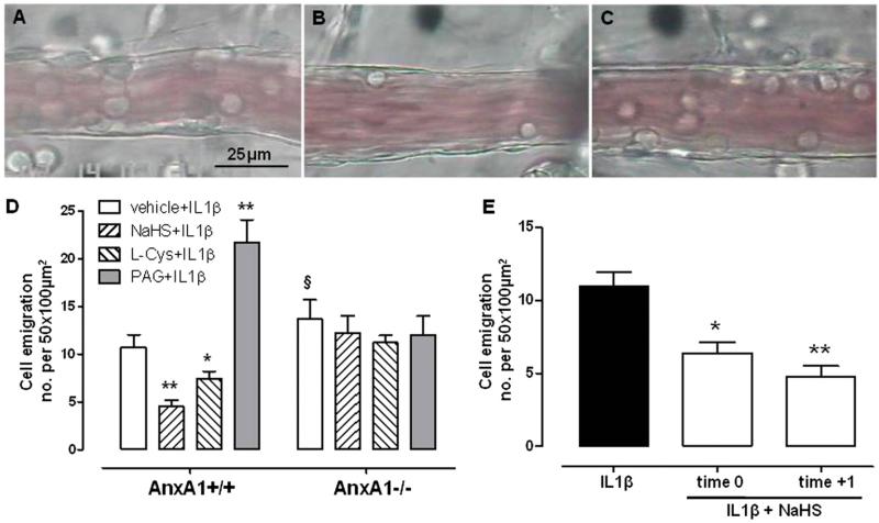
Fig. 3.
Intravital microscopy analysis of postcapillary venules in AnxA1+/+ or AnxA1−/− mice showing emigrated leukocytes. (A) Representative picture of postcapillary venule in AnxA1+/+ animal treated with IL-1β (10 ng/mouse i.p., 2 hours). (B) Representative picture of postcapillary venule in AnxA1+/+ animal treated with IL-1β (10 ng/mouse i.p., 2 hours), where NaHS (100 μmol/kg s.c.) was given 1 hour before IL-1β injection. (C) Representative picture of postcapillary venule in AnxA1−/− animal treated with IL-1β (10 ng/mouse i.p., 2 hours), where NaHS (100 μmol/kg s.c.) was given 1 hour before IL-1β injection. Mice were pretreated with vehicle, NaHS (100 μmol/kg s.c.), or l-cysteine (L-cys; 1000 μmol/kg s.c.) 1 hour before stimulation with IL-1β (10 ng/mouse i.p., 2 hours), and the number of adherent leukocytes was analyzed (expressed as number of cells per 50 × 100 μm2). (D) Pretreatment with PAG (10 mg/kg i.p.) was performed 30 minutes before IL-1β injection. (E) Number of emigrated leukocytes in AnxA1+/+ animal treated with IL-1β (10 ng/mouse i.p., 2 hours), where NaHS (100 μmol/kg s.c.) was administered at the same time as IL-1β (time 0) or 1 hour after its injection (time +1). Statistical analysis was made using two-way analysis of variance (*P < 0.05, **P < 0.01 versus vehicle; §P < 0.05 versus AnxA1+/+; n = 6).

