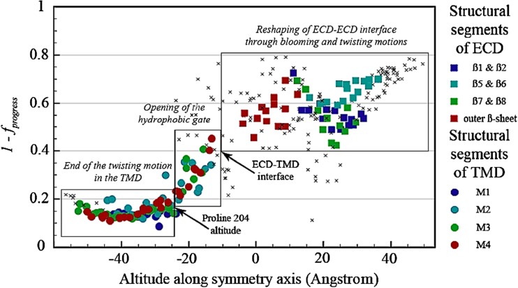
Fig. 4.
Coarsed grained molecular dynamics trajectories for the gating of the ion channel of the G. violaceus receptor. Opening of the ion channel is from right to left. The iENM web server (http://enm.lobos.nih.gov) was used to generate plausible trajectories between the four closed GLIC pH 7, the open GLIC pH 4 and the locally closed structures. The trajectories are mapped onto ECD and TMD reaction coordinates to quantify the differential progress of conformational change in these two domains. Residues belonging to structural segments (loops) are displayed with colored symbols (see color code on the right); all other residues are displayed with small crosses. The figure illustrates the stepwise progress of the concerted conformational change: from right to left channel opening process starting at the ECD, from left to right channel closing process starting at the TMD. From Sauguet et al. 2014a, Figure S18c.

