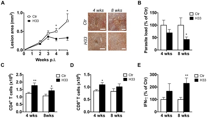
Figure 4. Blocking JAM-C improves the Th1 cell response and favours healing in C57BL/6 mice.
(A–E) Mice were inoculated with L. major in the ear dermis and treated with H33 or isotype control antibody for 3 weeks, twice a week. (A) The area of the lesion was monitored weekly and representative pictures of ear lesions are shown at 4 and 8 weeks p.i. Scale bars, 0.5 mm. Data represent the mean ± SEM of twenty mice per group pooled from two separate experiments for the time point 4 weeks; and fifteen mice per group pooled from two separate experiments for the time point 8 weeks. (B) The parasite burden in infected ears was measured by LDA 4 and 8 weeks p.i. Data are expressed as a percentage of the mean of the control group ± SEM of mice from panel A. (C–D) The number of draining lymph node CD4+ (C) and CD8+ (D) T cells analyzed by flow cytometry 4 and 8 weeks p.i. Data represent the mean ± SEM of mice from panel A. (E) Draining lymph node cells were restimulated for 72 hrs with UV-irradiated L. major and the secreted IFN-γ was measured. Data are expressed as a percentage of the mean of the control group ± SEM of mice from panel A. Data were analyzed by the unpaired Student's t test with *:p<0.05 and **: p<0.01. For panels B and E, raw data from one experiment are also provided in S8 Figure.

