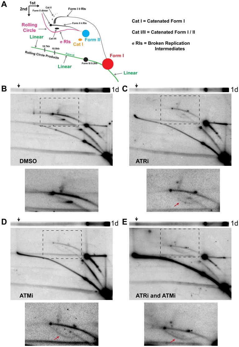
Figure 2. ATM or ATR inhibition increases broken replication forks and linear viral DNA replication products.
(A) Diagram of 2D gel electrophoresis of undigested circular dsDNA [86]. (B, C, D, E) Southern blots of the first dimension of a neutral 1D gel (top panel) or 2D gel (middle panel) from SV40-infected BSC40 cells exposed to DMSO (B), ATRi (C), ATMi (D), or ATRi and ATMi (E) during the last 28 h of a 48 h SV40 infection. Arrow on 1D gel points to the location of the ∼20 kb replication product. Bottom panel: Enlargement of the picture within the boxed area in middle panel. Exposure of the bottom panel was increased to enhance visualization of θ and σ replication intermediates shown in (A). Arrow in lower panels points to location of the σ arc.

