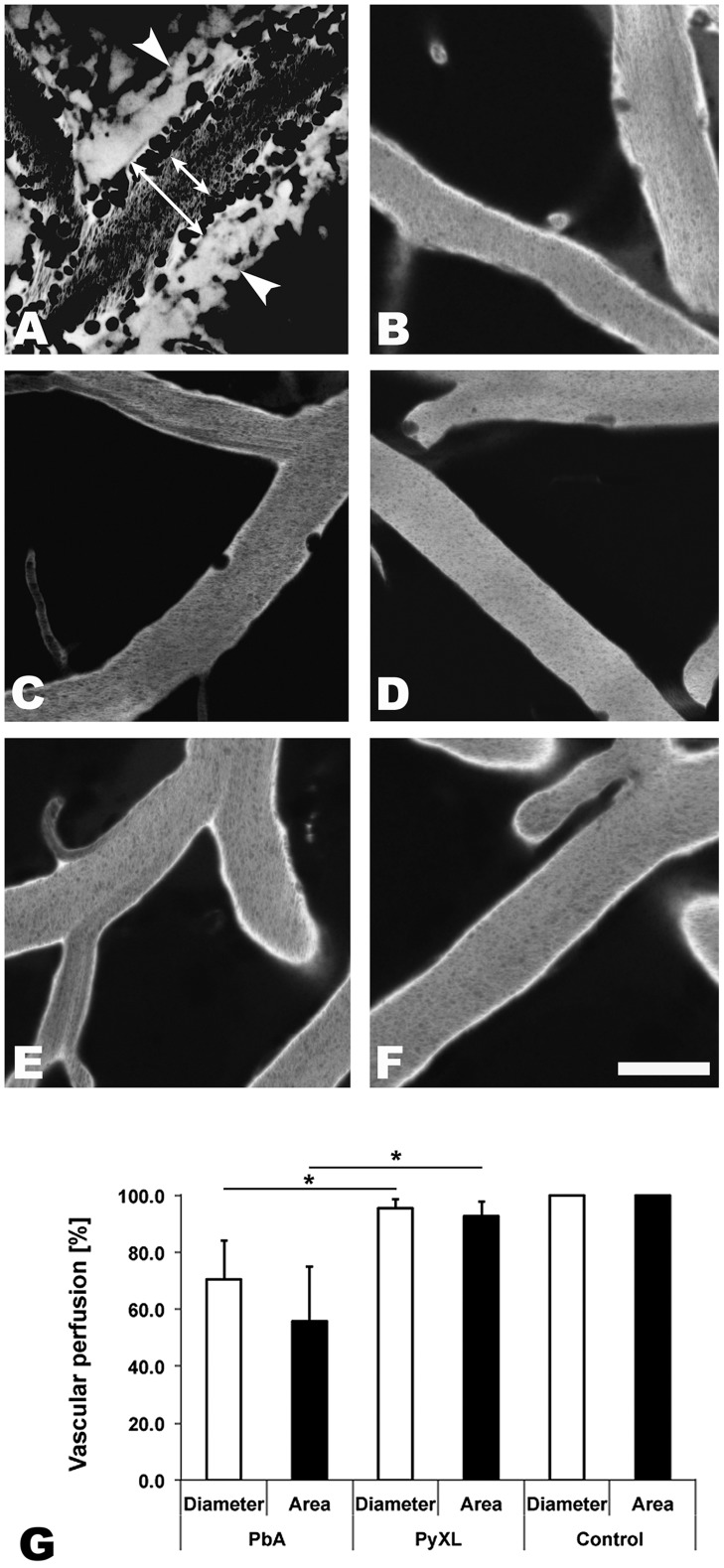
Figure 1. ECM correlates with microrheological alterations in postcapillary venules.
CBA/CaJ mice were infected with PbA, PyXL, or no parasites. To assess the blood flow within the cortical microvasculature, time sequences were converted to minimal projections. A) In mice with ECM, the functional postcapillary venule diameter (short arrow), i.e. the perfused portion of the vessel, is considerably reduced compared to the entire vessel diameter (long arrow). Visualization of the vascular lumen with Evans blue reveals a zone along the endothelium of postcapillary venules (A) that contains adherent leukocytes (dark circles or ovals), but is devoid of RBC (dark streaks in the center). Note that migrating leukocytes are represented multiple times in minimal projections. Leakage of Evans blue into the perivascular space and brain parenchyma is apparent on either side of the postcapillary venule (arrowheads). B) Arterioles from mice with symptomatic ECM do not exhibit any restriction in diameter. C) The small number of adherent leukocytes (dark circles) in mice with hyperparasitemia does not cause any significant restriction in the functional postcapillary venule lumen. Neither arterioles from mice with hyperparasitemia (D) nor postcapillary venules or arterioles from uninfected control mice (E and F) exhibit any microrheological alterations. Scale bars = 20 µm. See Videos S1–S5 for the corresponding dynamic data. G) Measurement of the total and functional vascular diameters and cross-sections reveals that the blood flow in postcapillary venules from mice with ECM, but not from mice with hyperparasitemia, is severely restricted. Postcapillary venules from uninfected control mice exhibit no luminal restriction.

