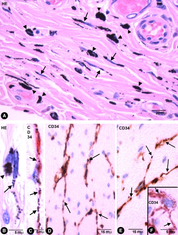
Fig. 1.

Stromal cells in enteric wall tattooed with India ink. (A) Characteristics of stromal cells (arrows) and macrophages (arrow-heads) in the submucosa with engulfed pigmented particles in an haematoxylin and eosin stained section. Note that the intracellular pigment in some stromal cells shows a linear distribution drawing their processes. (B) Detail of a stromal cell (arrow) and a macrophage (arrowhead) with endocytozed particles. (C) A bipolar CD34+ phTC with intracellular pigment (arrows) in submucosa. (D–F) bipolar and Multipolar CD34+ phTCs with intracellular pigment (arrows) between SMCs of muscular propria (D and E) and myenteric plexus ganglia (F).
