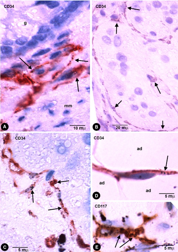
Fig. 3.

Enteric wall tattooed with India ink. (A) CD34+ phTCs around the basal portion of a gland (g) and lining the mucosa surface of muscularis mucosae (mm). (B and C) CD34+ phTCs (arrows) arranged around myenteric plexus ganglia. (D) A CD34+ phTC (arrow) between adipocytes (ad). (E) An occasional CD117 (c-kit) stained interstitial cell of Cajal (ICC) with endocytozed India ink particles (arrows).
