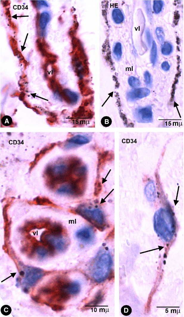
Fig. 5.

Stromal cells with intracytoplasmic pigmented particles in periodontal tissues with amalgam pigmentation. (A and C) CD34+ stromal cells with endocytozed particles (arrows). Note the arrangement of CD34+ stromal cells around different-sized vessels. (B) Stromal cells with abundant pigment around the media layer of a vessel in an HE-stained section. Note similar arrangement to CD34+ stromal cells in A. (D) A bipolar CD34+ stromal cell in the dermal interstitial compartment. Vl, Vessel lumen; ml, Media layer.
