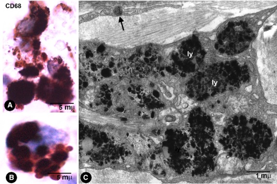
Fig. 7.

Macrophage characteristics. (A and B) CD68+ macrophages with abundant thick granules of India ink pigment in the intestinal wall. (C) Ultrastructural characteristics of macrophages in a pigmented melanocytic naevus. Note the abundant particles in lysosomes (ly). In the upper part of the image, a telopode of a phTC with an endocytozed melanosome (arrow).
