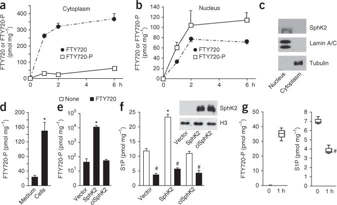
Figure 1.

FTY720-P is produced in the nucleus by SphK2. (a–d) SH-SY5Y neuroblastoma cells were treated with 5 μM FTY720 for the indicated times (a,b) or for 6 h (d) (N = 3 independent cell cultures per group). Cytoplasmic (a) and nuclear (b) levels of FTY720 and FTY720-P were determined by liquid chromatography–electrospray injection–tandem mass spectroscopy (LC-ESI-MS/MS). (c) Equal amounts of protein were separated by SDS-PAGE and immunoblotted with SphK2-specific antibody. Antibodies against lamin A/C and tubulin were used as nuclear and cytosolic markers, respectively. (d) Total intracellular and secreted FTY720-P were determined by LC-ESI-MS/MS. *P < 0.001 as compared to medium; unpaired Student’s t-test. (e,f) SH-SY5Y neuroblastoma cells transfected with vector, SphK2 or catalytically inactive SphK2G212E (ciSphK2) constructs were treated without or with FTY720 for 6 h and nuclear levels of FTY720-P (e) and S1P (f) determined by LC-ESI-MS/MS. Equal expression was confirmed by immunoblotting. Data are expressed as mean ± s.d. *P < 0.005 as compared to vector; #P < 0.001 as compared to untreated; unpaired Student’s t-test. All western blots were performed three times. Full-length blots are presented in Supplementary Figure 10. (g) Hippocampal neurons were treated with FTY720 for 1 h and nuclear levels of FTY720-P and S1P determined by LC-ESI-MS/MS (N = 3 independent cell culture replicates per group). Box plots indicate median (center line), 25–75th percentile (box limits), and minimum and maximum (whiskers).
