
Figure 7.
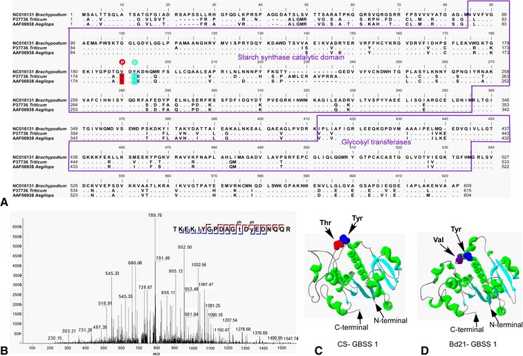
Phosphorylation of GBSSI. A, Amino acid sequence alignment of granule-bound starch synthases (GBSSI proteins). The phosphorylated residues are marked. B, The mass spectrometric spectrum of the phosphopeptide. C, 3D structure is shown for GBSSI of Chinese Spring (CS; common wheat) and Aegilops peregrina. D, 3D structure is shown for GBSSI of Brachypodium distachyon Bd21.