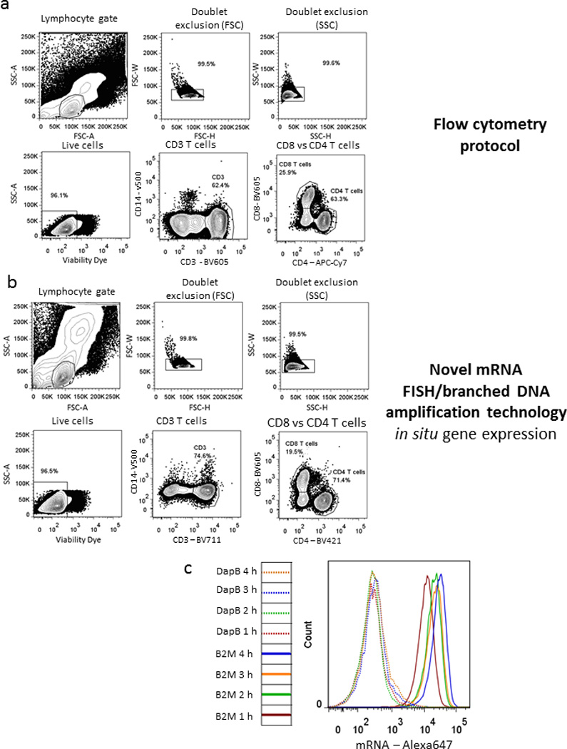
Figure 1. Antibody staining and detection of housekeeping genes.

Comparison of physical and phenotypic characteristics of PBMCs stained with a dead cell dye and fluorescent antibodies for CD3, CD4, CD8 and CD14 using either (a) a standard surface stain flow cytometry protocol or (b) the new flow-RNA assay (representative out of 5). (c) Kinetic experiment of different hybridization incubation times following staining with either an irrelevant probe DapB or the housekeeping gene β2M (representative example out of 3).
