Figure 12.
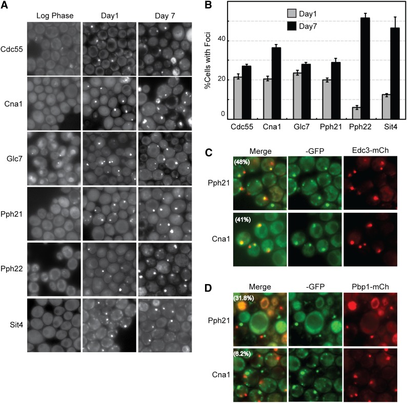
Particular S. cerevisiae protein phosphatases are also recruited to cytoplasmic foci in stationary-phase cells. (A) Cells expressing the indicated protein phosphatase-GFP fusions were examined by fluorescence microscopy during log phase and after 1 or 7 days of growth in rich medium. (B) Quantitation of the microscopy data shown in A. (C and D) The colocalization of the protein phosphatase subunits Pph21 and Cna1 with a reporter for P-bodies (C, Edc3-mCh) or stress granules (D, Pbp1-mCh). The extent of colocalization is indicated by the percentage value in parentheses in the merged images.

