Figure 7.
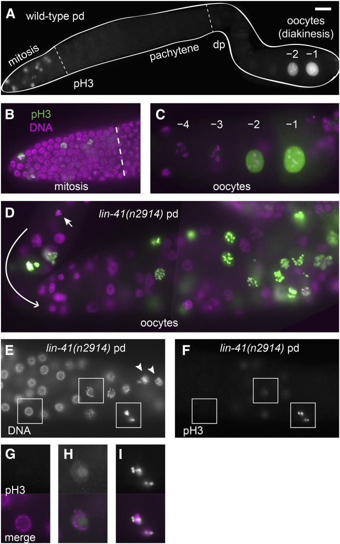
Distinct patterns of phospho-histone H3 accumulation in the wild-type and lin-41 mutants. (A–C) In wild-type animals, antibodies detecting phosphorylated histone H3 (pH3) strongly stain some of the nuclei in the distal mitotic region and also stain the nuclei of the most proximal oocytes (–1 and –2), but do not stain pachytene or diplotene (dp) nuclei (A). The overlap of pH3 (green) with a nuclear DNA stain (DAPI, magenta) is shown at higher magnification in the mitotic region (B) and in the last few oocytes (C). (D–I) In lin-41(n2914) animals, many oocyte nuclei accumulate pH3, including nuclei near the loop (D, curved arrow; the earliest nucleus accumulating pH3 on chromosomes is indicated by a short arrow). The first pH3-positive chromosomes are found just after lin-41(n2914) germ cells exit from pachytene (E and F). Arrowheads in E indicate nuclei with decondensed chromosomes, which are located proximal to the first pH3-positive nuclei. Nuclei in boxes are magnified two-fold in G–I and include a typical pachytene-stage nucleus with no pH3 accumulation (G), a later pachytene-stage nucleus with faint nuclear pH3 (H), and condensed pH3-positive chromatin immediately after pachytene (I). Bar, 20 μm (A); 10 μm (B–F); and 5 μm (G–I).

