Figure 2.

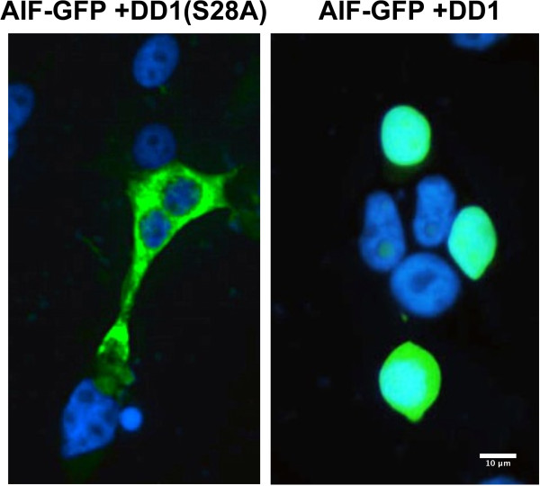
DD1-mediated apoptosis in LNCaP cells leads to changes in mitochondrial membrane permeability and release of AIF which translocates to the cell nucleus. LNCaP-AI cells were first transfected with a plasmid to express AIF-GFP. Twenty-four h later cells were transfected to express DD1 which leads to apoptosis, or DD1(S28A) which is inactive [DD1 and DD1(S28A) were expressed as GAL4 fusion proteins to ensure nuclear localization]. Fifteen h after the second transfection cells were fixed and permeablized for AIF-GFP fluorescence (green). Nuclei were stained with DAPI (blue). The Figure shows an extra-nuclear mitochondrial distribution of AIF-GFP in cells expressing DD1(S28A) while in cells expressing DD1, AIF-GFP is localized to the nucleus.
