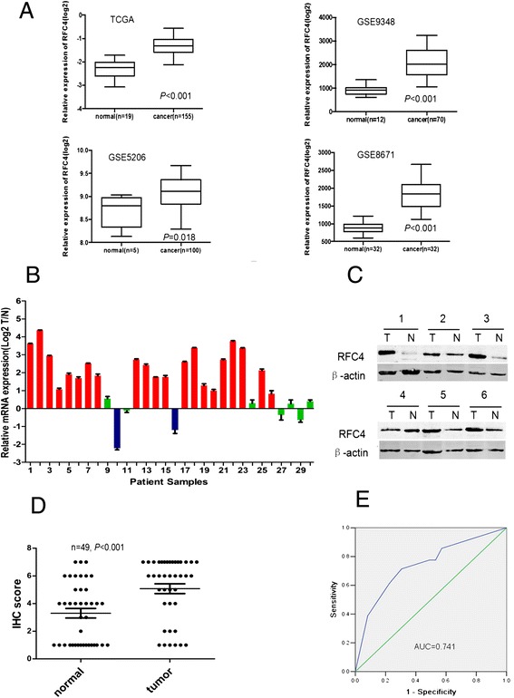
Figure 1.

RFC4 is overexpressed in CRC tissues. (A) RFC4 mRNA levels were assessed from The Cancer Genome Atlas (TCGA) and the Gene Expression Omnibus databases (GEO, GSE9348, GSE5206 and GSE8671). Bounds of the boxes denote interquartile range; solid lines denote median, whiskers denote 95% and 5% confidence limits, respectively. (B) Expression of RFC4 mRNA in 30 paired CRC tumor and adjacent non-tumor tissues. Each bar is the log2 value of the ratio of RFC4 expression levels between CRC (T) and matched normal tissues (N) from the same patient. Red bars (bar value ≥1) indicate high RFC4 mRNA levels. Blue and green bars represent low and normal expression of RFC4 mRNA, respectively. Each sample was analyzed twice. (C) Western blots for RFC4 in matched-pairs of CRC tumor and non-tumor tissues. High RFC4 protein expression in CRC is seen in 12 of 16 patients. (D) Distribution of RFC4 immunoreactivity scores in tumor and paired-non-tumor tissues from the tissue microarray. CRC tissues exhibite higher RFC4 expression than normal colonic tissues. (E) ROC curves analysis to determine the cutoff score for the high expression of RFC4. The area under curve (AUC) is 0.741, and the cutoff point of RFC4 for overexpression is 4.5.
