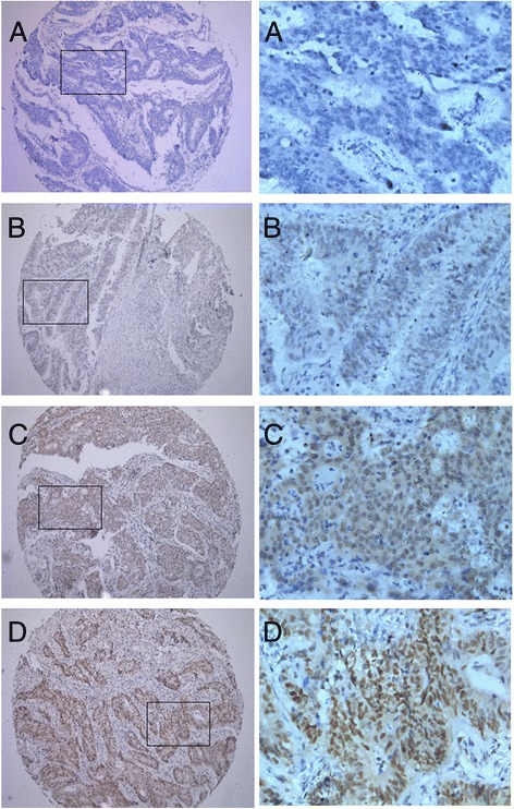
Figure 2.

Expression pattern of RFC4 in CRC by immunohistochemistry. The low power photomicrographs (left side) in each section show the full tissue spot (×100), while the high power photomicrographs (right side) show details of RFC4 expression (×400). Nuclear staining is considered RFC4 expression positive. (A) negative nuclear staining; (B) weak nuclear staining; (C) moderate nuclear staining; (D) strong nuclear staining.
