Figure 7.
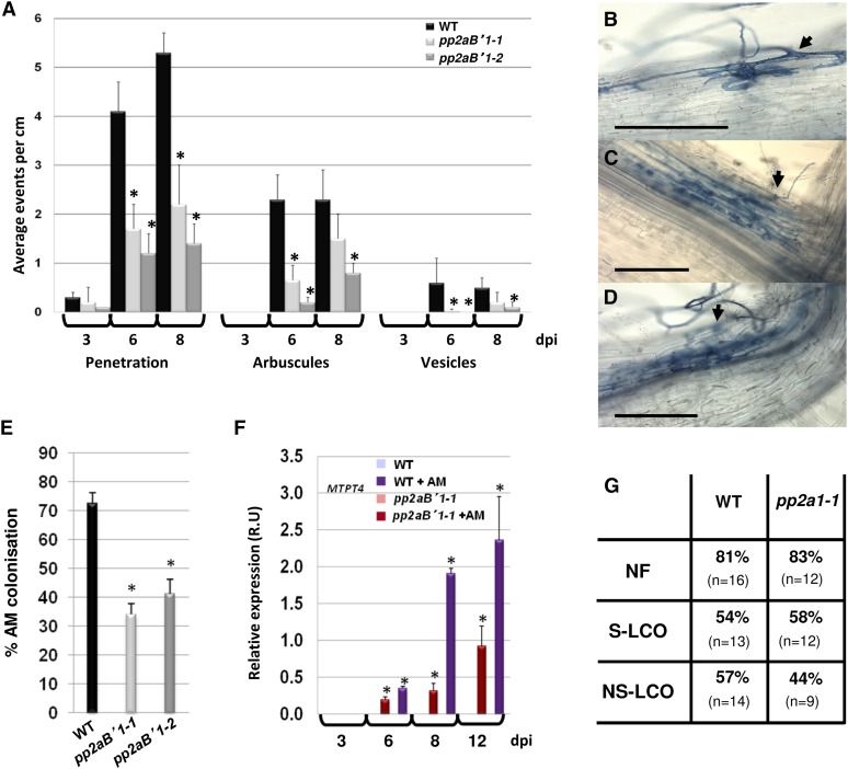
PP2AB′1 is required for AM penetration and propagation. A to E, AM propagation analysis 3, 6, 8, and 12 dpi with germinating R. irregularis spores infecting wild-type (WT) and pp2aB′1 mutant roots. A, The numbers of penetration events with intraradical mycelium (B), penetration events with cortical cells containing arbuscules (C), and penetration events with cortical cells containing arbuscules and vesicles (D; Supplemental Fig. S10) were quantified per cm of root. B to D, Bright-field images show the propagation of the fungus infecting the pp2aB′1-1 mutant 6 dpi. Black arrows show the fungal penetration. Bars = 200 μm. The AM fungus is ink stained. E, Percentage of AM root length colonization with R. irregularis germinating spores 12 dpi. F, Quantitative RT-PCR to monitor MtPT4 expression in wild-type M. truncatula roots infected with R. irregularis (WT + AM) and in pp2aB′1-1 during fungal propagation (A–E). The expression is normalized to EF-1α level. Values are means ± se from three biological replicates. Asterisks indicate significant differences from the wild type (A and E) or from the control noninoculated roots (the wild type and pp2aB′1-1) in F (Student’s t test; P ≤ 0.01; n = 24). G, Calcium spiking measurements in the wild type and pp2aB′1-1 mutants in response to 10−9 m nodulation factor (NF), 10−8 m sulfated lipochitooligosaccharides (S-LCO), and 10−6 m NS-LCO. The percentage of cells spiking is indicated, and n = number of cells recorded.

