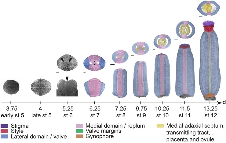
Figure 1.
Arabidopsis gynoecium development. Transmitted light confocal images of the remaining floral meristem (stage [st] 5) and the first stages of gynoecial primordia development (stages 6 and 7), and false-colored DIC images of floral stages 8 to 12 gynoecia along a developmental time scale showing the time in days after floral initiation at the end of each stage. Early and late stage 5 as well as upper images at floral stages 6 and 7 are viewed from above. Lower floral stage 6 image is viewed from the lateral side. Lower images of floral stages 7 to 12 gynoecia are viewed from the medial side. Upper images of floral stages 8 to 12 gynoecia show transverse sections. Stages and time scale are adapted after Smyth et al. (1990) and Sessions (1997). White dashed lines indicate lateral plane, black dashed lines indicate medial plane, arrowheads indicate lateral crease, and asterisks indicate CMM. Bars = 10 µm (stages 5–7), 25 µm (stages 8–10), and 50 µm (stages 11 and 12).

