Figure 5.
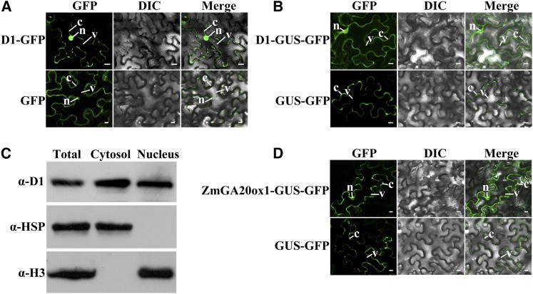
Subcellular localization of D1 protein. A, B, and D, D1-GFP fusion and GFP proteins (A), D1-GUS-GFP and GUS-GFP fusion proteins (B), and ZmGA20ox1-GUS-GFP (NP_001241783) and GUS-GFP fusion proteins (D) were transiently expressed in tobacco leaf epidermal cells and were analyzed by fluorescent laser confocal microscope. C, Western-blot detection of D1 protein in nucleus and cytosol fractions of maize seedlings. Total, Total proteins from shoot and root. The protein amount was loaded identically for each antibody. α-D1, Anti-D1 (ZmGA3ox2) antibody; α-HSP82, anti-heat shock protein 82 antibody (cytosolic marker); C, cytosol; DIC, Differential interference contrast; N, nucleus; V, vacuole. Bars = 10 μm.

