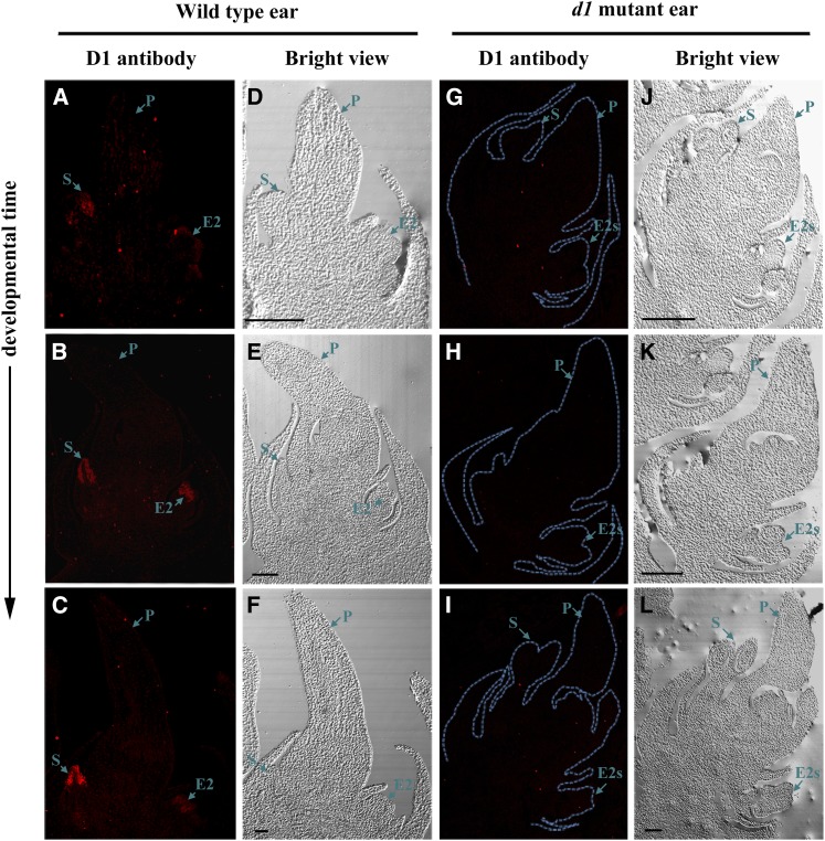
Figure 8.
Immunohistochemical detection of D1 protein in developing maize female florets. Sections of wild-type and d1 ears (1–1.5 cm) were incubated with anti-D1 antibody and followed with secondary antibody labeled with Alexa Fluor 594, which produces red fluorescent signals under a fluorescent microscope. A to C, Wild-type female florets incubated with D1 antibody. D to F, Bright views of A to C. G to I, d1 Female florets incubated with D1 antibody. J to L, Bright views of G to I. E2, Secondary ear florets; E2s, stamen of E2 florets; P, pistil of E1 florets; S, stamen of E1 florets. Bars = 200 μm.

