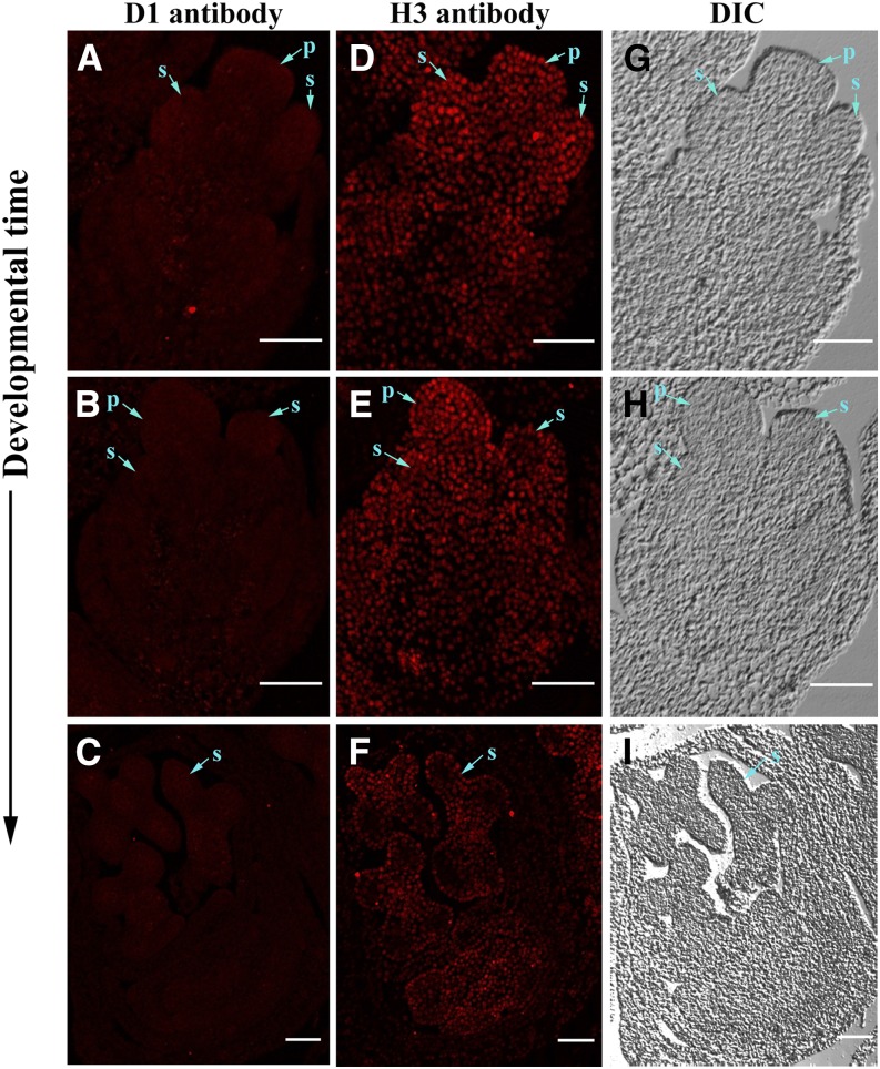
Figure 9.
Immunohistochemical detection of D1 protein in developing maize male florets. Sections of wild-type tassels from 1 to 1.5 cm were incubated with anti-D1 antibody and anti-H3 antibody followed with secondary antibody labeled with Alexa Fluor 594, which produces red fluorescent signals under fluorescent microscope. A to C, Wild-type male florets incubated with D1 antibody. D to F, Wild-type male florets incubated with H3 antibody. G to I, Bright views of A to C and D to F. Developmental time arrow points to late stage. DIC, Differential interference contrast; P, pistil; S, stamen. Bars, 100 μm.

