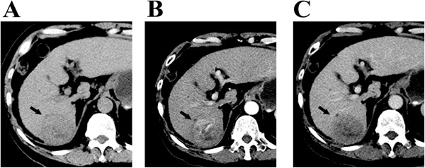
Figure 1.

Contrast-enhanced CT images obtained in a patient with 6-cm single HCC before TACE treatment. (A-C) show a patient with hepatitis B-induced liver cirrhosis and a 6-cm solitary HCC tumor in the hepatic segment VI. The contrast-enhanced CT scan before TACE revealed arterial enhancement of the HCC lesion.
