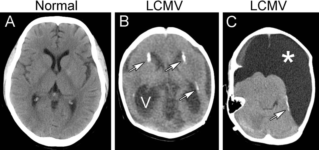
Figure 2.
Neuroimaging studies reveal neuropathology induced by congenital LCMV infection. Shown here are MRI scans from normal children (left column) and children with congenital LCMV infection (right column). A. In the midsagittal plane of a normal child, the cerebellum (arrow) is large and fills the posterior fossa. B. In congenital LCMV infection, the virus can impair cerebellar growth and lead to cerebellar hypoplasia (arrow). C. In the horizontal plane of a normal child, the cerebral cortex is folded into a complex set of gyri and sulci (arrow). D. In congenital LCMV infection, the cerebral cortex is often featureless and smooth, lacking normal gyri and sulci, and reflecting a neuronal migration disturbance (arrows). This patient also has periventricular calcifications (arrowheads) and ventriculomegaly.

