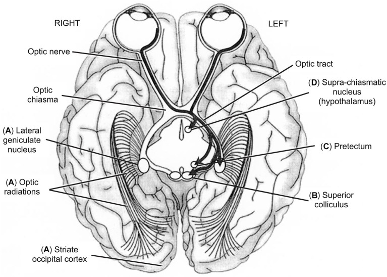
Figure 4.
Inferior view of the brain showing the five main visual pathways, in which left is right and vice-versa. (A) Retino-occipital or retino-cortical visual pathway. (B) Retino-collicular or retinotectal visual pathway. (C) Retino-pretectal visual pathway. (D) Retino-hypothalamic visual pathway. Accessory optic visual pathway is not shown. Thick arrows show the different trajectories of ganglion neurons originating in leftward hemi-retinas (temporal hemi-retina of the left eye and nasal hemi-retina of the right eye). Ganglion neurons originating in rightward hemi-retinas exhibit mirror trajectories but are not illustrated here for clarity of display. From O.A. Coubard (© O.A. Coubard, with permission).

