Fig. 3.
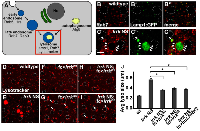
Abnormally expanded late endosomal and lysosomal compartments in lrrk NS flies. (A) Schematic depicting the endosomal and autophagy pathways, and highlighting markers for different pathway compartments. The lysosomal compartment, indicated by the red box, is the focus of this figure. Nu, nucleus. (B,C) Antibody staining for the late endosomal protein Rab7 in follicle cells from stage-12 wild-type (B) and lrrk NS (C) egg chambers shows dramatically enlarged Rab7-positive compartments in lrrk NS. These enlarged Rab7-positive compartments accumulate the lysosomal marker Lamp1:GFP (C′ vs B′). Arrowheads indicate Rab7 staining of the late endosomal membrane. (D–I) Staining of lysosomes with the acidophilic dye Lysotracker in the indicated genotypes shows dramatically enlarged lysosomes in lrrk NS follicle cells (E) relative to wild type (D); this phenotype can be rescued by follicle-cell-specific expression of wild-type lrrk (H). In an otherwise wild-type background, expression of lrrkGS (G), but not wild-type lrrk (F), results in perinuclear clustering of lysosomes. In lrrk NS flies, expression of lrrkGS suppresses the enlarged lysosome phenotype; however, perinuclear clustering of lysosomes is still observed (I). Arrowheads indicate perinuclear lysosome clusters. (J) Quantification of average lysosome size in the indicated genotypes as determined by Lysotracker staining of follicle cells from stage-12 egg chambers. Whereas lrrk NS results in a dramatic increase in average lysosome size, this phenotype is significantly suppressed by follicle-cell-specific expression of wild-type lrrk, lrrkGS or human LRRK2. n=16, 34, 16, 16 and 17 egg chambers each, respectively, for the genotypes in J. *P<0.0001. lrrk NS is lrrk1/2. Follicle cell nuclei are outlined by dashed white lines in B–I. Scale bar: 5 μm.

