Fig. 1.

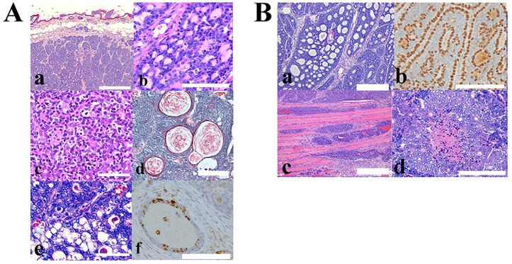
Representative histopathological findings of the dermal tumors that appeared in the C57BL/6-Tg (ITF-TMEM207) mouse. (A) Lobules of the basaloid cells arranged in jigsaw or mosaic pattern without connection to the epidermis. These morphological features are characteristic of human cylindromas and spiradenomas (a; scale bar: 1000 μm). (b) Hyaline matrix materials are regularly dispersed in basaloid tumor cells, which is a feature of cylindroma. Scale bar: 50 μm. (c) Trabecular tumor nests composed of small basaloid cells and large cuboidal cells with thin membranous stroma, which is often found in spiradenoma. Scale bar: 100 μm. Follicular (d; scale bar: 500 μm) and sebaceous differentiations (e; scale bar: 100 μm) were partially observed in several tumors. Note the tumor nests with keratin-filled cysts that are found in trichoepithelioma (d). Adipophilin immunoreactivity also indicated sebaceous differentiation (f; scale bar: 50 μm). (B) Cutaneous adnexal tumors with malignant phenotypes were also observed. Cribriform architecture was found in the tumor nest (a). The p63 immunoreactivity was detected in the peripheral tumor cells of the nest, and myoepithelial-like cells faced pseudoglands in the cribriform lesions (b). These features were compatible with those of adenoid cystic carcinomas. Several tumors partially exhibited the invasion to skeletal muscle (c) or comedo-type necrosis (d), which is known to be malignant characteristics of cutaneous adnexal tumors. (Ba–d) Scale bars: 100 μm.
