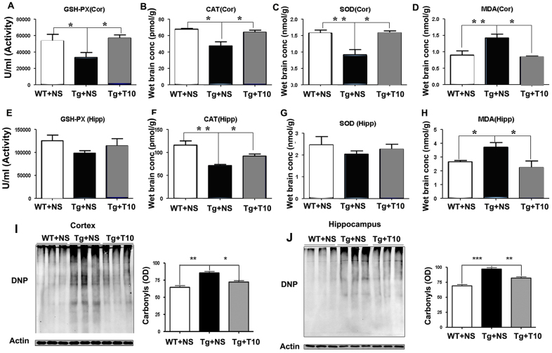
Fig. 7.
Treatment with triptolide had anti-oxidative effects on the brains of 5XFAD mice. (A,E) ELISA was used to detect the activity of GSH-Px in the cortex (Cor) and hippocampus (Hipp) of saline-treated wild-type mice (WT+NS), saline-treated 5XFAD mice (Tg+NS) and triptolide (T10)-treated 5XFAD mice (Tg+T10). (B,F) CAT levels in the cortex and hippocampus of the three groups of mice were measured by using ELISA. (C,G) SOD contents of the cortex and hippocampus of the three groups of mice. (D,H) The production of MDA in the cortex and hippocampus of the three groups of mice. (I,J) The protein samples of cortex or hippocampus were treated with dinitrophenyl hydrazine and subjected to western blotting. The levels of oxidative proteins were determined by using densitometry to quantify positive bands of 2,4-dinitrophenyl (DNP)-modified proteins. Actin was used as an internal control (n=6 animals per group), *P<0.05, **P<0.01, ***P<0.001 versus Tg+NS, one-way ANOVA with Tukey’s post hoc test. Conc, concentration; OD, optical density.

