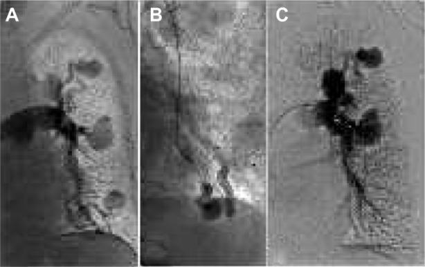
Figure 1.

Pulmonary arteriovenous-fistula treatment.
Notes: (A) Angiography reveals multiple left pulmonary arteriovenous malformations with shunts in a patient with Rendu–Osler–Weber disease. (B) After placement of a diagnostic catheter in the arterial feeder of the shunt, embolization is performed with an AVP4 device that can be observed in fluoroscopy. (C) Final angiography shows complete occlusion of the three distal arteriovenous shunts.
Abbreviation: AVP, Amplatzer vascular plug.
