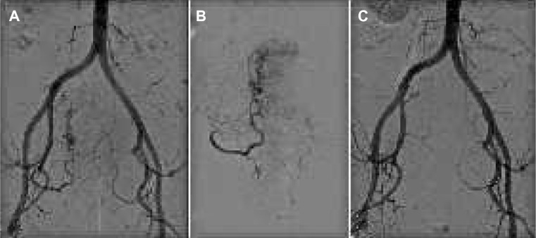
Figure 4.

Uterine fibroid treatment.
Notes: (A) Pelvic angiogram shows tortuous and dilated uterine arteries feeding a huge uterine fibroid with its typical corkscrew appearance. (B) Selective angiography demonstrates a hypervascular uterine fibroid before embolization. The first outgoing branch of the uterine artery supplies proper blood flow to the cervix uteri, and should be preserved during embolization. (C) After embolization with polyvinyl alcohol particles, the main uterine artery is still patent but with decreased blood flow and devascularization of the abnormal fibroid vessels.
