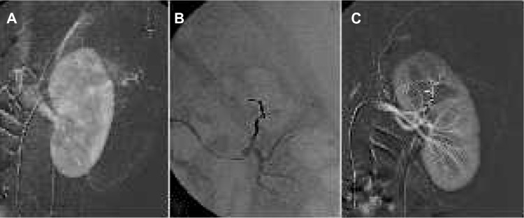
Figure 5.

Renal angiomyolipoma treatment.
Notes: (A) Selective renal angiogram shows abnormal blood vessels at the upper part of the kidney. (B) After embolization of the feeding vessel through a microcatheter and using Onyx, the embolization cast appears radiopaque on fluoroscopy. (C) Final angiogram confirms complete embolization of the angiomyolipoma, preserving the renal parenchyma.
